OCR · GCSE · Biology
Course version: J247 · Version from Sept 2018
Use of appropriate apparatus, techniques and magnification, including…
Full specification wording
Microscopy: Use of appropriate apparatus, techniques and magnification, including microscopes, to make observations of biological specimens and produce labelled scientific drawings
Learners prepare temporary mounts: a single layer of onion epidermis stained with iodine, or cheek epithelium stained with methylene blue, is placed on a clean slide and a coverslip is lowered at…
- Point 1 of 2
- 3 key terms
Study notes
From the teaching notes for this specification point.
The idea
Learners prepare temporary mounts: a single layer of onion epidermis stained with iodine, or cheek epithelium stained with methylene blue, is placed on a clean slide and a coverslip is lowered at about 45 degrees with a mounted needle to avoid trapping air bubbles. The slide is viewed on a compound light microscope, starting on the lowest power objective (4x), focusing with the coarse knob then the fine knob, before moving to higher power. Biological drawings use unbroken pencil lines, no shading, accurate proportions, and state the total magnification (eyepiece x objective, e.g. 10 x 40 = 400x). Labels are straight, uncrossed lines touching the structure named, such as nucleus, cell wall and cytoplasm.
What earns the marks
- Lower the coverslip slowly at an angle using a mounted needle so air bubbles are not trapped over the stained specimen.
- Begin viewing on the lowest power objective lens, focus with the coarse knob until the specimen appears, then sharpen with the fine knob before changing objective.
- Produce biological drawings with single unbroken pencil lines, no shading or cross-hatching, accurate proportions, and the total magnification stated.
- Draw straight, uncrossed label lines that touch the structure named, without arrowheads, and label only structures actually visible.
- Calculate total magnification by multiplying eyepiece magnification by objective magnification, e.g. 10 x 40 = 400x.
Common mistakes and how to put them right
Using a thick tissue sample that overlaps several cell layers, so light transmission is poor and cellular detail is obscured.
Put it right by peeling a single thin layer such as onion epidermis.
Drawing with sketchy lines, artistic shading, or omitting the stated magnification.
Put it right by using one continuous pencil line per outline and recording the total magnification.
Focusing with the high-power objective first, which risks cracking the coverslip and damaging the lens.
Put it right by focusing on low power, then moving to high power and using only the fine knob.
Exam tips
- Calculate total magnification by multiplying the eyepiece lens magnification (typically 10x) by the objective lens magnification.
- When asked to evaluate a biological drawing in an exam, check for uncrossed label lines, absence of shading, and clear continuous outlines.
Try the lesson
The start of the Microscopy: Use of appropriate apparatus, techniques and magnification, including microscopes, to make observations of biological specimens and produce labelled scientific drawings lesson, made from teaching notes for this specification point.
Have a go first
Which piece of apparatus is used to view a biological specimen at high magnification?
Listen
Learners prepare temporary mounts: a single layer of onion epidermis stained with iodine, or cheek epithelium stained with methylene blue, is placed on a clean slide and a coverslip is lowered at about 45 degrees with a mounted needle to avoid trapping air bubbles. The slide is viewed on a compound light microscope, starting on the lowest power objective (4x), focusing with the coarse knob then the fine knob, before moving to higher power. Biological drawings use unbroken pencil lines, no shading, accurate proportions, and state the total magnification (eyepiece x objective, e.g. 10 x 40 = 400x). Labels are straight, uncrossed lines touching the structure named, such as nucleus, cell wall and cytoplasm. A strong answer earns marks for these points. Lower the coverslip slowly at an angle using a mounted needle so air bubbles are not trapped over the stained specimen. Begin viewing on the lowest power objective lens, focus with the coarse knob until the specimen appears, then sharpen with the fine knob before changing objective. Produce biological drawings with single unbroken pencil lines, no shading or cross-hatching, accurate proportions, and the total magnification stated. Draw straight, uncrossed label lines that touch the structure named, without arrowheads, and label only structures actually visible. A common slip: Using a thick tissue sample that overlaps several cell layers, so light transmission is poor and cellular detail is obscured. Instead, peeling a single thin layer such as onion epidermis. Exam tip: Calculate total magnification by multiplying the eyepiece lens magnification (typically 10x) by the objective lens magnification.
See it
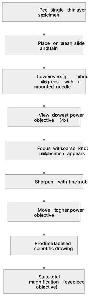
Key words
Say what each word means before you turn the card.
microscope operation and focus
Try answering aloud or in your head before turning the card.
scientific drawing conventions
Try answering aloud or in your head before turning the card.
cellular magnification
Try answering aloud or in your head before turning the card.
See every definition
- microscope operation and focus
- The slide is viewed on a compound light microscope, starting on the lowest power objective, focusing with the coarse knob then the fine knob, before moving to higher power.
- scientific drawing conventions
- Biological drawings use unbroken pencil lines, no shading, accurate proportions, and state the total magnification, with straight, uncrossed label lines touching the structure named.
- cellular magnification
- Total magnification is the eyepiece magnification multiplied by the objective magnification, for example 10 x 40 = 400x.
Label the diagram
Some boxes have lost their names. Pick the right name for each number.
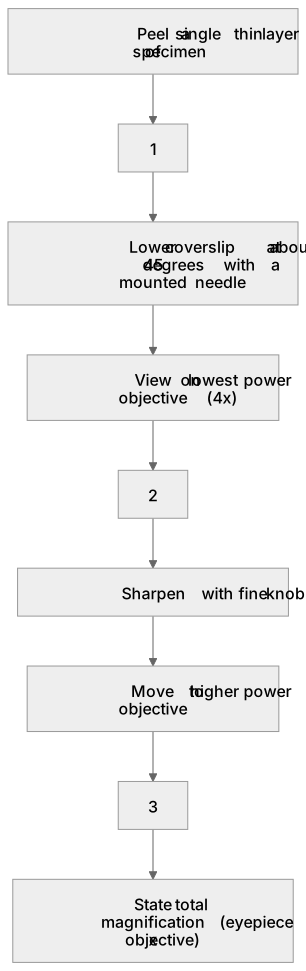
1Box 1
2Box 2
3Box 3
There is more to this lesson
The full lesson goes on to check your understanding and help you earn the marks. Create a free account to carry on, with your progress saved.
A week of Premium from your first lesson · Then the free plan continues · No card, no charge
Test your understanding of Microscopy: Use of appropriate apparatus, techniques and magnification, including microscopes, to make observations of biological specimens and produce labelled scientific drawings
Active recall beats re-reading. Take an instant quiz now to test what you’ve learned and lock it into long-term memory.
A week of Premium from your first lesson · Then the free plan continues · No card, no charge