Pearson Edexcel · GCSE · Combined Science
Exchange and transport in animals
This topic explains how and why animals move essential substances around their bodies. You'll master the critical relationship between size and exchange efficiency, and discover how the heart, lungs, and blood vessels are perfectly adapted for their life-saving roles.
- 6 min read
- 3 worked examples
- 5 practice questions
- 6 key terms
Study Notes

Overview
Welcome to Topic 8: Exchange and Transport in Animals. This is a foundational topic in Combined Science because it explains the physiological mechanisms that keep multicellular organisms alive. While a single-celled organism can rely on simple diffusion to obtain oxygen and remove waste, larger animals require complex, highly adapted systems to survive.
In this topic, you will explore the mathematical concept of Surface Area to Volume Ratio (SA:V) and see exactly why size dictates the need for lungs and a heart. You will examine the structural adaptations of the alveoli, the different types of blood vessels, and the anatomy of the double circulatory system. Examiners frequently test this topic by asking you to link a specific structural adaptation to its function, or by asking you to calculate cardiac output. Mastering this topic also provides crucial synoptic links to respiration and cellular biology.
Listen to our comprehensive revision podcast to solidify your understanding:
Key Concepts
Concept 1: Surface Area to Volume Ratio (SA:V)
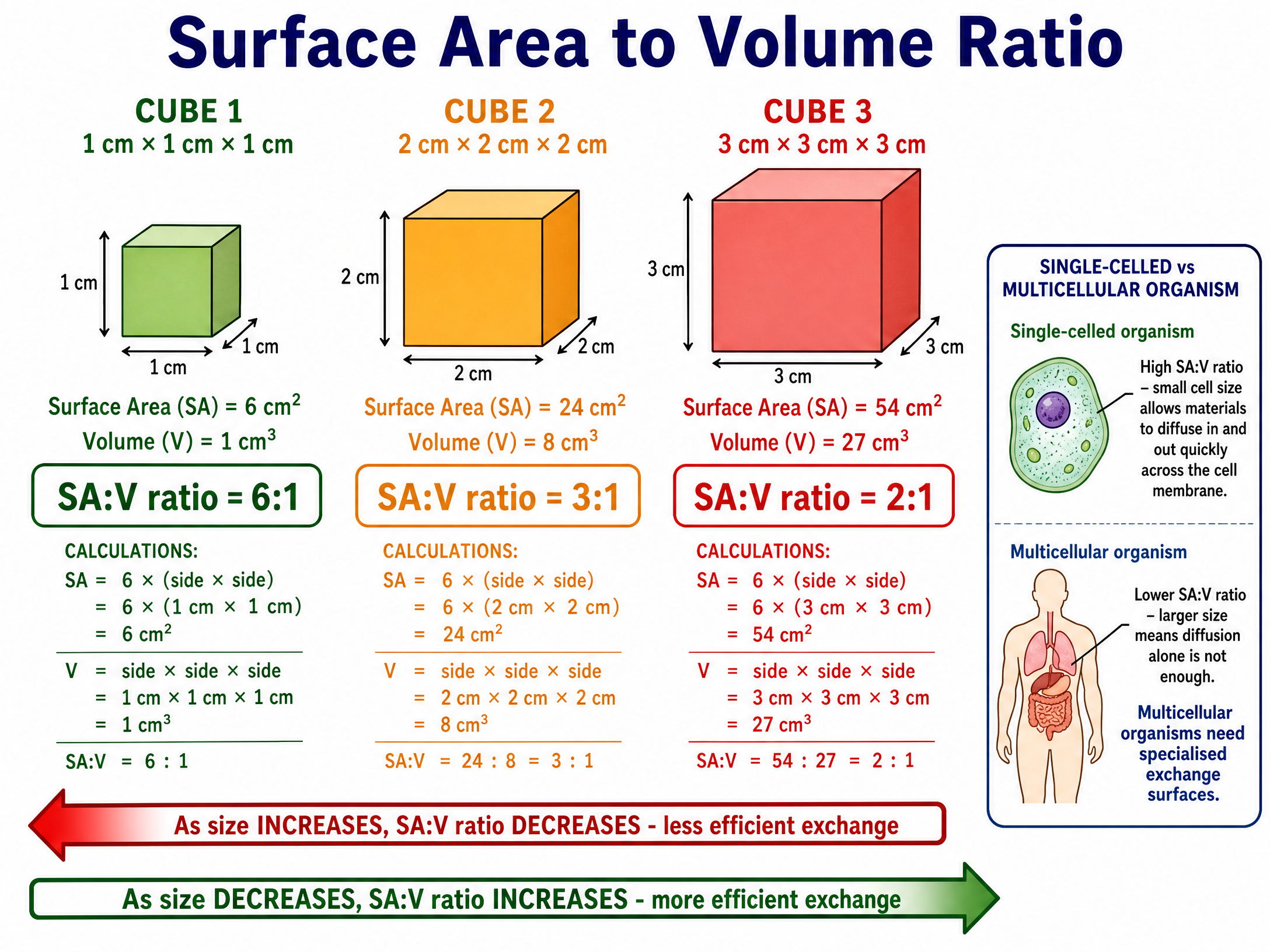
As an organism increases in size, its volume grows at a much faster rate than its surface area. This means that a large multicellular organism has a relatively small surface area compared to its massive volume. Consequently, simple diffusion across the outer surface is far too slow to supply all the internal cells with oxygen and nutrients, or to remove toxic waste products like carbon dioxide.
To overcome this, multicellular organisms have evolved specialised exchange surfaces (like the lungs) and transport systems (like the circulatory system) to move substances efficiently between the exchange surface and the body cells.

Concept 2: Adaptations of the Alveoli
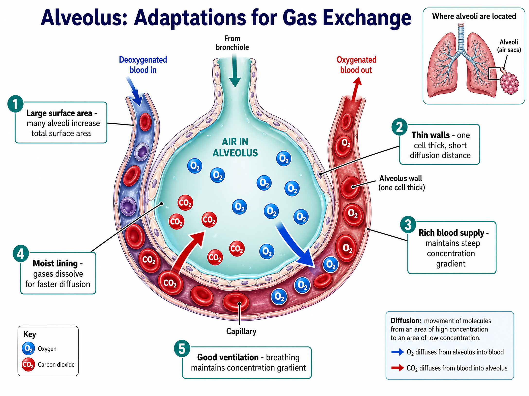
The lungs contain millions of tiny air sacs called alveoli, where gas exchange occurs. Oxygen diffuses from the air in the alveoli into the blood, while carbon dioxide diffuses from the blood into the alveoli to be exhaled. The alveoli are perfectly adapted to maximise the rate of diffusion:
- Large Surface Area: The millions of alveoli provide a massive total surface area, allowing more molecules to diffuse simultaneously.
- Thin Walls: The alveolar walls and capillary walls are both just one cell thick. This provides a very short diffusion distance, significantly increasing the rate of diffusion.
- Rich Blood Supply: A dense network of capillaries surrounds each alveolus. Blood is constantly flowing, bringing carbon dioxide and taking away oxygen. This maintains a steep concentration gradient, which is essential for rapid diffusion.
- Good Ventilation: Breathing constantly refreshes the air in the alveoli, ensuring oxygen concentration remains high and carbon dioxide concentration remains low, further maintaining the steep concentration gradient.

Concept 3: Blood Vessels
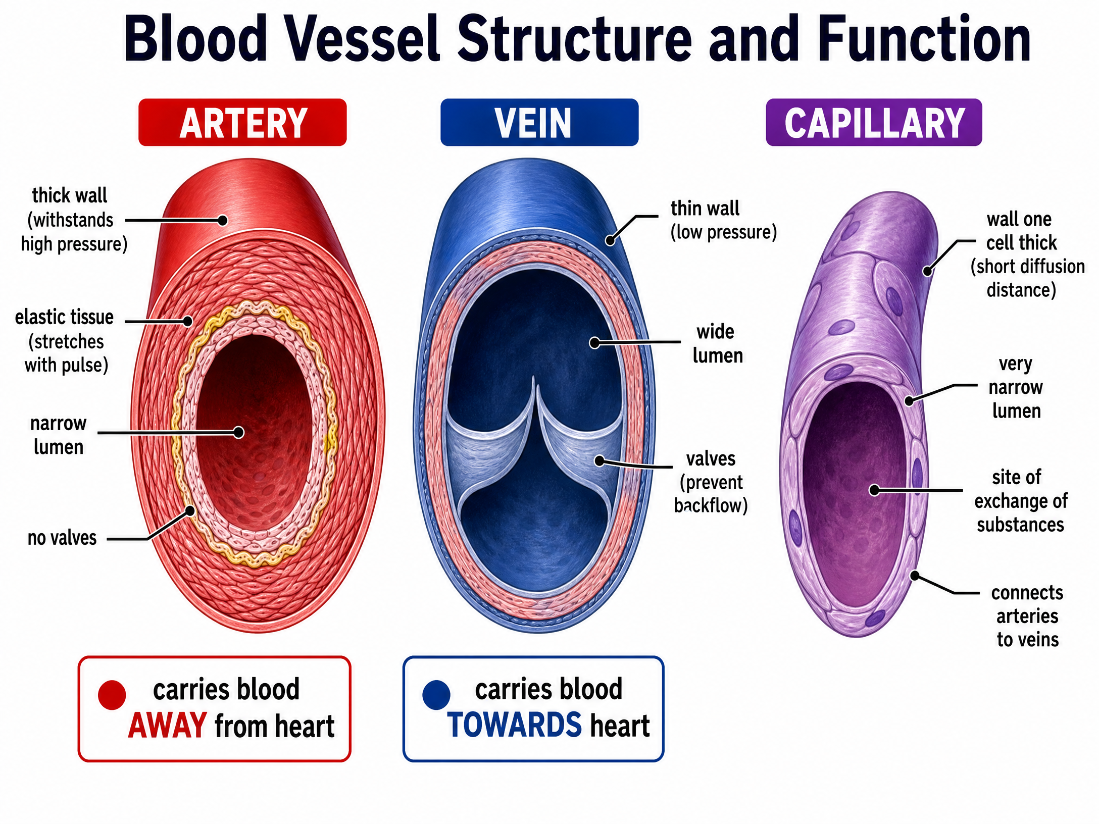
The circulatory system uses three main types of blood vessels, each structurally adapted to its specific function:
- Arteries: Carry blood away from the heart. Because the blood is pumped directly from the heart, it is at very high pressure. Arteries have thick, muscular, and elastic walls to withstand this pressure and stretch with each pulse. They have a relatively narrow lumen (the central hole).
- Veins: Carry blood towards the heart. The blood is at a much lower pressure here. Veins have thinner walls and a wider lumen than arteries. Crucially, veins contain valves to prevent the backflow of blood, ensuring it flows in only one direction.
- Capillaries: These are the microscopic vessels where the actual exchange of substances (oxygen, glucose, carbon dioxide) takes place between the blood and the body tissues. Their walls are only one cell thick, providing a very short diffusion distance.

Concept 4: The Heart and the Double Circulatory System
Humans have a double circulatory system, meaning blood passes through the heart twice for every complete circuit of the body. The right side of the heart pumps deoxygenated blood to the lungs, while the left side pumps oxygenated blood to the rest of the body. This system ensures blood reaches the body tissues at a high pressure, delivering oxygen quickly.
The heart has four chambers: the right atrium, right ventricle, left atrium, and left ventricle. The walls of the ventricles are much thicker than the atria because they have to pump blood out of the heart. Specifically, the left ventricle has a much thicker muscular wall than the right ventricle. This is a common exam question! It is thicker because the left ventricle must pump blood all the way around the entire body (requiring high pressure and force), whereas the right ventricle only pumps blood to the nearby lungs.
Mathematical/Scientific Relationships
Surface Area to Volume Ratio
- Formula:
Ratio = Surface Area / Volume - Usage: Used to demonstrate why larger organisms need transport systems. Always simplify the ratio (e.g., 24:8 becomes 3:1).
- Must memorise: Yes.
Cardiac Output
- Formula:
Cardiac Output = Stroke Volume × Heart Rate - Symbols:
- Cardiac Output: Total volume of blood pumped by the heart per minute (usually in cm³/min or L/min).
- Stroke Volume: Volume of blood pumped out of the heart with each beat (usually in cm³).
- Heart Rate: Number of heartbeats per minute (bpm).
- Usage: Used to calculate heart efficiency. Pay close attention to units!
- Must memorise: Yes, this is rarely given on the formula sheet.
Practical Applications
Understanding cardiac output and blood vessel structure is fundamental in medicine. For instance, measuring heart rate and stroke volume helps cardiologists diagnose heart failure. Knowledge of the coronary arteries (which supply the heart muscle itself with oxygen) is vital for treating coronary heart disease with stents or bypass surgery.
Visual Resources
3 diagrams and illustrations
Interactive Diagrams
2 interactive diagrams to visualise key concepts
Conceptual Flow Outline
Flowchart showing the path of blood through the double circulatory system.
Conceptual Flow Outline
The direction of gas diffusion driven by concentration gradients in the lungs.
Worked Examples
3 worked examples — open one to explore the question and available guidance.
Practice Questions
Test your understanding — click to reveal model answers
Compare the structure of an artery with the structure of a vein. (4 marks)
Hint: Think about the wall thickness, the size of the hole in the middle, and what might be inside the vessel.
Explain how the alveoli are adapted for efficient gas exchange. (6 marks)
Hint: You need to give at least three structural features and explain exactly how each one speeds up diffusion.
A marathon runner has a resting heart rate of 50 bpm and a resting stroke volume of 100 cm³. Calculate their resting cardiac output in dm³/min. (3 marks)
Hint: Remember that 1 dm³ is the same as 1 Litre, which is 1000 cm³.
Explain why the left ventricle has a thicker muscular wall than the right ventricle. (2 marks)
Hint: Think about where each side of the heart is pumping blood to.
Explain why single-celled organisms do not need a complex circulatory system. (3 marks)
Hint: Think about the SA:V ratio and the distance substances need to travel.