Pearson Edexcel · GCSE · Combined Science
Key concepts in biology
Master the fundamental building blocks of life. This topic covers cell structure, specialised cells, and the microscopy techniques used to discover them—essential knowledge that underpins the entire biology specification and guarantees marks in your exams.
- 7 min read
- 3 worked examples
- 5 practice questions
- 6 key terms
Study Notes
Overview

Welcome to Key Concepts in Biology, the foundation of your GCSE Combined Science course. Every living organism is made of cells, and understanding their structure is critical because it connects to almost every other biological topic you will study, from respiration and photosynthesis to genetics and disease.
In this topic, you will explore the differences between complex eukaryotic cells (like ours) and simpler prokaryotic cells (like bacteria). You will learn to identify sub-cellular structures (organelles) and, crucially, explain how their structure allows them to carry out specific functions. Examiners frequently test this topic through labeling diagrams, comparing cell types, and calculating magnification from microscope images.
Listen to the audio guide below for a comprehensive review of this topic, complete with an exam-style quick-fire quiz:
Key Concepts
Concept 1: Eukaryotic and Prokaryotic Cells
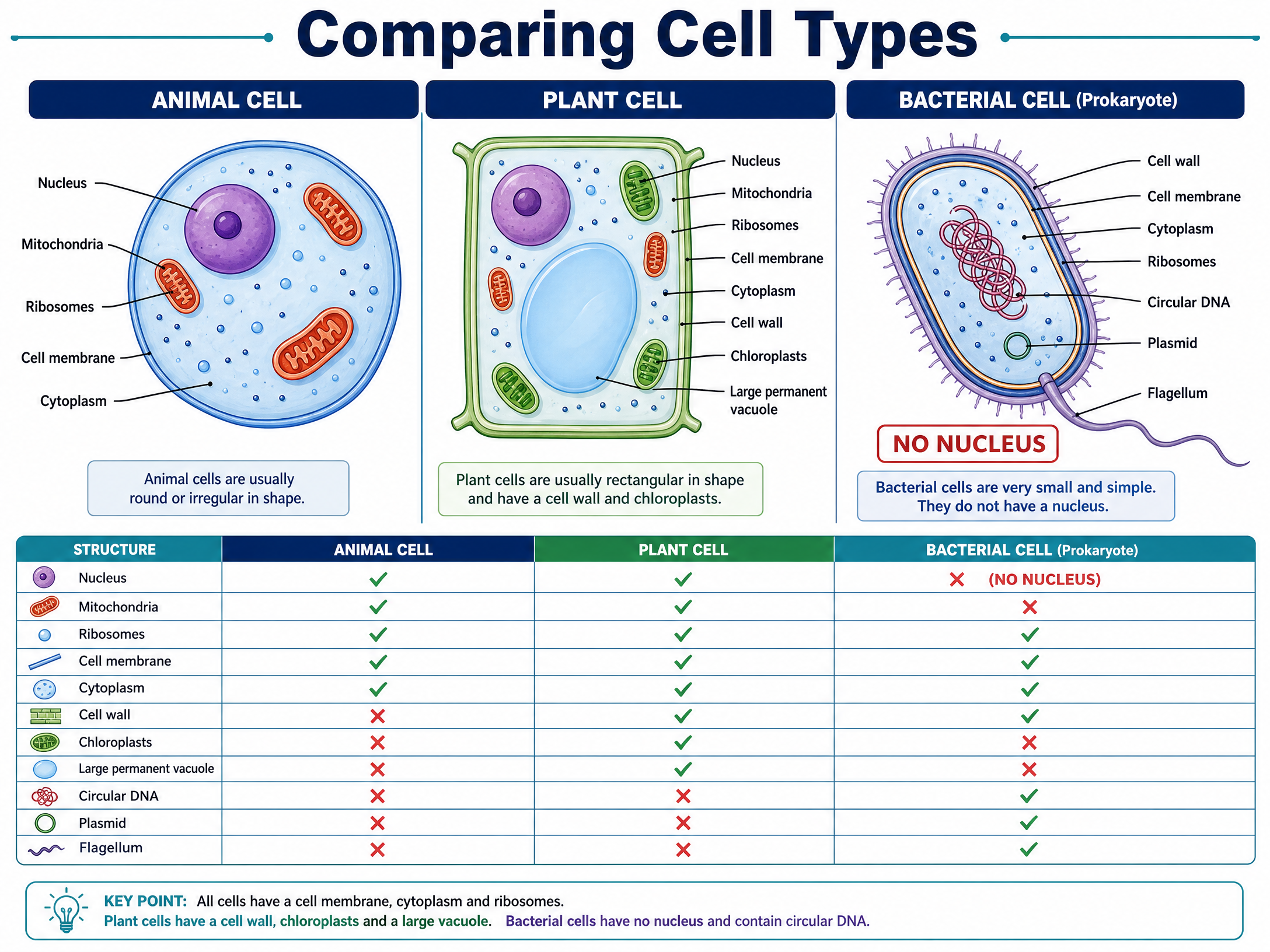
All cells fall into two broad categories: eukaryotic and prokaryotic.
Eukaryotic cells are complex and include all animal and plant cells. Their defining feature is that their genetic material (DNA) is enclosed within a nucleus. Think of the nucleus as a secure vault protecting the cell's instruction manual.
Prokaryotic cells, such as bacteria, are much smaller and simpler. They do not have a nucleus. Instead, their genetic material floats freely in the cytoplasm as a single circular strand of DNA. They may also contain small rings of extra DNA called plasmids.
Example: If an exam question asks you to identify a bacterial cell from a diagram, look for the absence of a nucleus and the presence of a circular DNA strand or plasmids.
Concept 2: Sub-cellular Structures (Organelles)
To earn high marks, you must know the function of key organelles and which cell types contain them.

Structures in both Animal and Plant Cells:
- Nucleus: Contains genetic material that controls the activities of the cell.
- Cytoplasm: A gel-like substance where most chemical reactions happen. It contains enzymes that control these reactions.
- Cell Membrane: Holds the cell together and controls what goes in and out (selectively permeable).
- Mitochondria: These are the powerhouses of the cell. They are the site of most of the reactions for aerobic respiration, releasing energy that the cell needs to function.
- Ribosomes: The site of protein synthesis (where proteins are made).
Structures ONLY in Plant Cells:
- Rigid Cell Wall: Made of cellulose. It supports the cell and strengthens it.
- Permanent Vacuole: Contains cell sap (a weak solution of sugar and salts). It helps maintain the cell's turgor pressure.
- Chloroplasts: The site of photosynthesis, which makes food for the plant. They contain a green substance called chlorophyll, which absorbs the light needed for photosynthesis.
Concept 3: Cell Specialisation
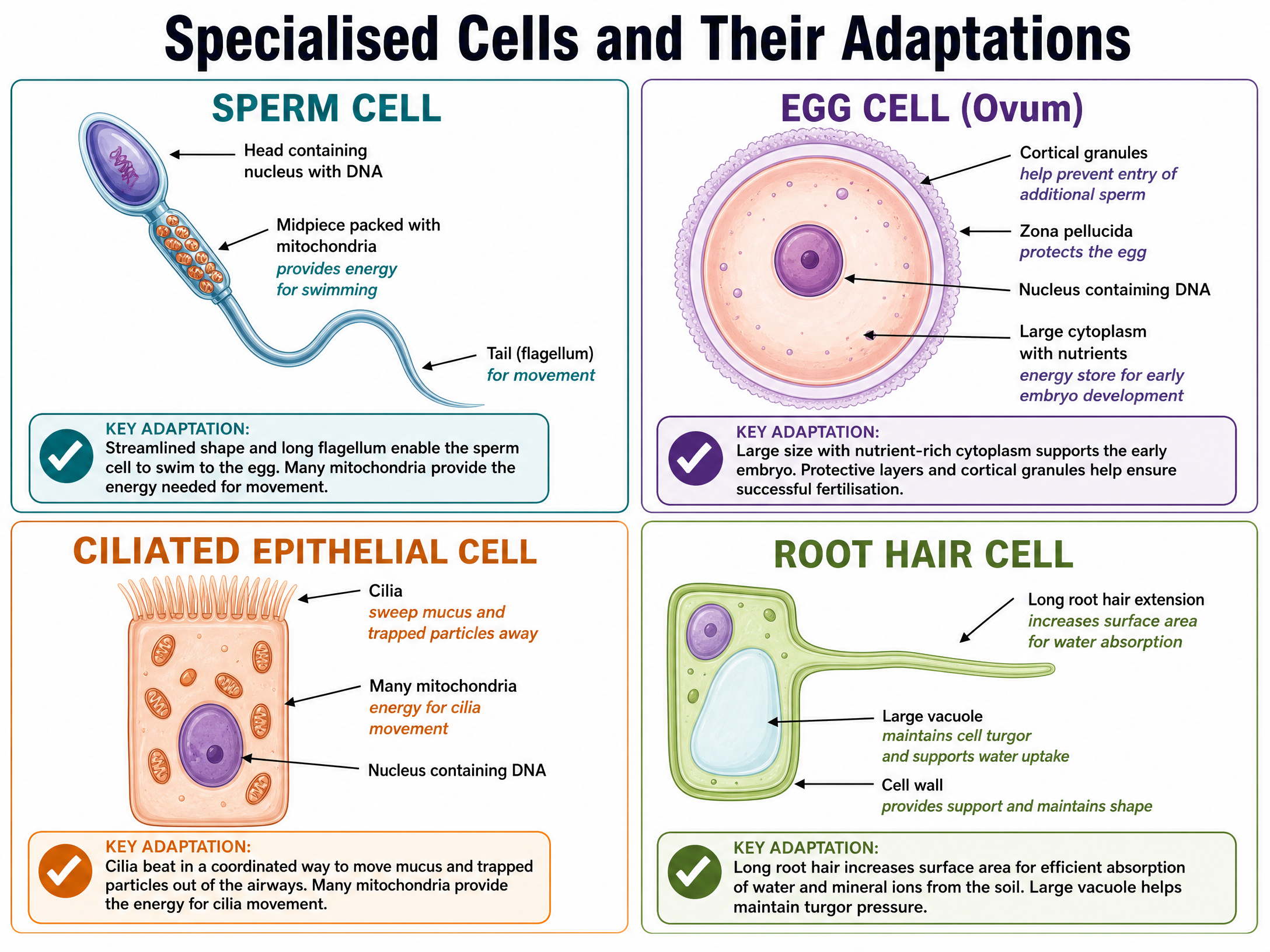
Cells differentiate to become specialised for a specific function. The structure of a specialised cell is perfectly adapted to its role. Examiner Tip: Always use the phrase 'so that' or 'because' to link the structural adaptation to the function.

- Sperm Cells (Reproduction): Adapted to transport the male DNA to the female DNA. It has a long tail (flagellum) and a streamlined head to help it swim. It contains lots of mitochondria to provide the energy needed for swimming. It also carries enzymes in its head to digest through the egg cell membrane.
- Nerve Cells (Rapid Signalling): Adapted to carry electrical signals from one part of the body to another. They are long to cover more distance and have branched connections at their ends to connect to other nerve cells and form a network throughout the body.
- Muscle Cells (Contraction): Adapted to contract quickly. They are long (giving them space to contract) and contain lots of mitochondria to generate the energy needed for contraction.
- Root Hair Cells (Absorbing Water and Minerals): Cells on the surface of plant roots which grow into long "hairs" that stick out into the soil. This gives the plant a large surface area for absorbing water and mineral ions from the soil. They do not contain chloroplasts as they are underground.
- Phloem and Xylem Cells (Transporting Substances): Form tubes which transport substances such as food and water around plants. Xylem cells are hollow in the centre, and phloem cells have very few subcellular structures, so that stuff can flow through them.
Concept 4: Microscopy
Our understanding of cells has been driven by advancements in microscope technology.
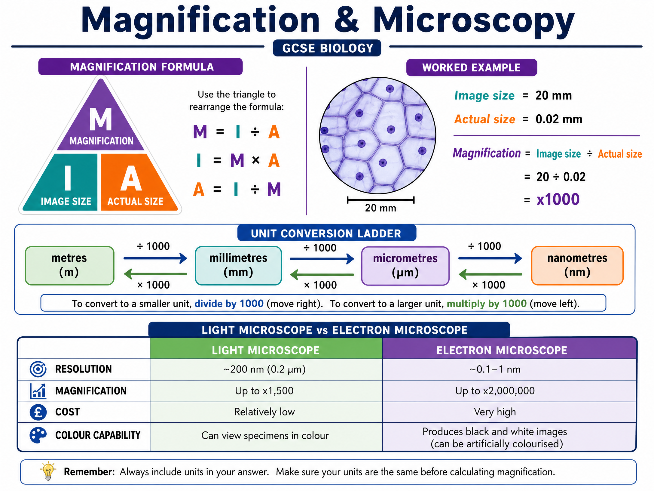
Light Microscopes use light and lenses to form an image of a specimen and magnify it. They let us see individual cells and large subcellular structures, like nuclei. They have a maximum magnification of around x1500 and a resolution of about 200nm.
Electron Microscopes use electrons instead of light to form an image. They have a much higher magnification (up to x2,000,000) and a much higher resolution (around 0.1nm) than light microscopes. This allows us to see much smaller things in more detail, like the internal structure of mitochondria and chloroplasts, and even tinier things like ribosomes and plasmids.
Mathematical/Scientific Relationships

The Magnification Formula
Magnification = Image Size ÷ Actual Size
- Magnification (M): How many times larger the image is than the real object (no units, just a number with 'x').
- Image Size (I): The size of the object as it appears in the drawing or photograph (usually measured with a ruler in mm).
- Actual Size (A): The real size of the object.
CRITICAL RULE: Before using the formula, you MUST ensure that the Image Size and Actual Size are in the same units.
Unit Conversions
Examiners will often give you measurements in different units to test your conversion skills.
- 1 millimetre (mm) = 1,000 micrometres (μm)
- 1 micrometre (μm) = 1,000 nanometres (nm)
To convert from mm to μm, multiply by 1000.
To convert from μm to mm, divide by 1000.
Practical Applications
Required Practical: Using a Light Microscope
You must know how to prepare a slide (e.g., onion epidermal tissue) and use a light microscope to observe it.
Key Steps:
- Add a drop of water to the middle of a clean slide.
- Cut up an onion and separate it out into layers. Use tweezers to peel off some epidermal tissue from the inner surface.
- Place the epidermal tissue into the water on the slide.
- Add a drop of iodine solution. Iodine is a stain used to highlight objects in a cell by adding colour to them.
- Place a cover slip on top. Stand the slip upright on the slide, next to the water droplet. Then carefully tilt and lower it so it covers the specimen. Try not to get any air bubbles under there, as they'll obstruct your view of the specimen.
Common Errors: Trapping air bubbles under the cover slip, or using too much stain which obscures the cells.
Visual Resources
3 diagrams and illustrations
Interactive Diagrams
2 interactive diagrams to visualise key concepts
Conceptual Flow Outline
Classification of cell types and their sub-cellular structures.
Conceptual Flow Outline
Unit conversion pathway for microscopy calculations.
Worked Examples
3 worked examples — open one to explore the question and available guidance.
Practice Questions
Test your understanding — click to reveal model answers
A student observes a palisade mesophyll cell under a light microscope. Name three structures they might see. (3 marks)
Hint: Palisade cells are plant cells found in leaves.
Explain why electron microscopes have been crucial in advancing our understanding of sub-cellular structures. (3 marks)
Hint: Think about what electron microscopes have that light microscopes lack, and what this allows scientists to see.
A bacterial cell has a length of 2 μm. Calculate its length in millimetres. Give your answer in standard form. (2 marks)
Hint: To convert from micrometres to millimetres, you need to divide.
A student measures the length of a mitochondrion on an electron micrograph as 15 mm. The actual length of the mitochondrion is 3 μm. Calculate the magnification of the image. (3 marks)
Hint: Make sure both measurements are in the same unit before calculating.
Root hair cells and palisade mesophyll cells are both plant cells, but they have different structures. Compare their structures and explain how these differences relate to their functions. (6 marks)
Hint: Identify where each cell is found in the plant, what its job is, and what organelles it needs (or doesn't need) to do that job.