Pearson Edexcel · GCSE · Combined Science
Radioactivity
Master the hazards of ionising radiation, the crucial distinction between contamination and irradiation, and the essential safety precautions used in medicine. This guide provides the exact terminology and cause-and-effect reasoning examiners look for in 6-mark questions.
- 5 min read
- 3 worked examples
- 5 practice questions
- 6 key terms
Study Notes
Overview

Welcome to Topic 6: Radioactivity. This topic is fundamentally about the interaction between unstable atomic nuclei and living tissue. In Combined Science, examiners aren't just testing your ability to recall facts; they want to see if you can apply physical principles to real-world safety scenarios, particularly in medical settings.
Understanding the hazards of ionising radiation is crucial because it forms the basis for all radiation protection protocols. You will frequently encounter questions asking you to distinguish between contamination and irradiation, or to evaluate the risks and precautions associated with different types of radiation. This topic connects strongly with your biology knowledge of cells and DNA, and examiners often reward synoptic thinking that bridges these disciplines.
Listen to our comprehensive revision podcast to solidify your understanding:
Key Concepts
Concept 1: The Ionising Nature of Radiation
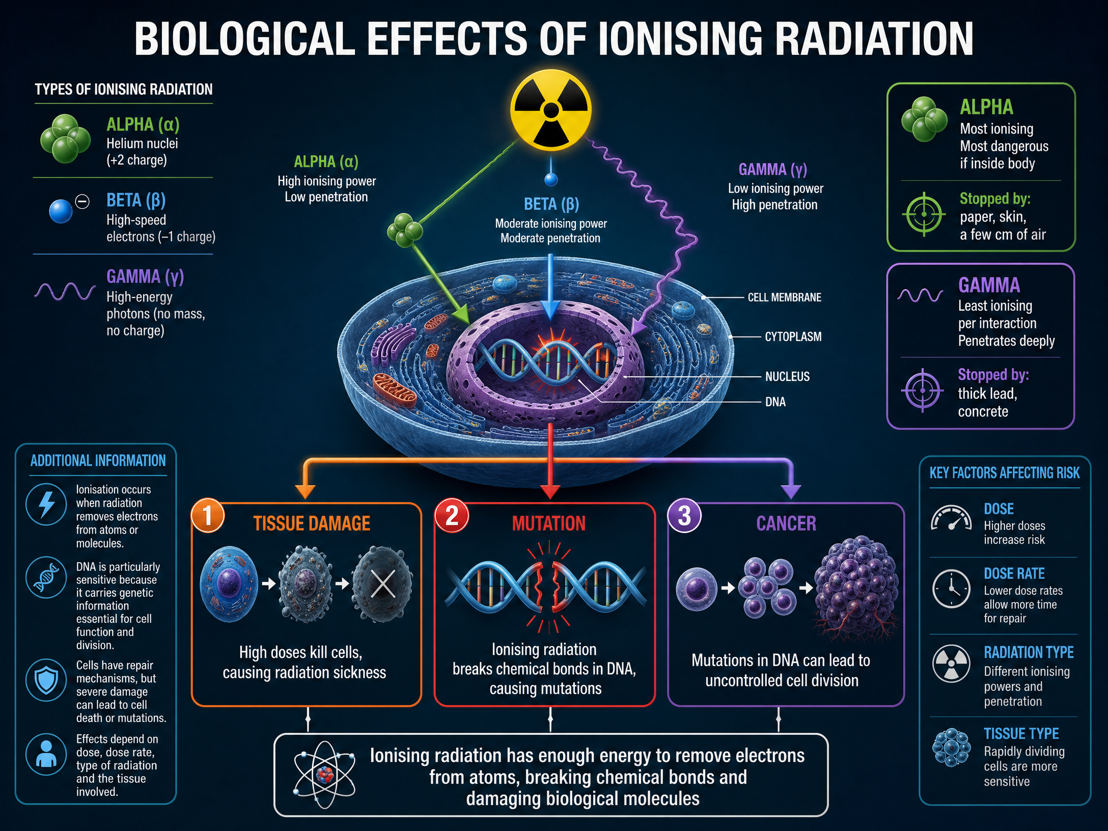
The fundamental reason radiation is hazardous is its ability to ionise atoms. Ionising radiation has enough energy to knock electrons out of the outer shells of atoms, turning neutral atoms into charged ions.
When this happens inside a living cell, it can break the chemical bonds holding complex biological molecules together. The most critical molecule in this context is DNA.
Example: If an alpha particle travels through a skin cell, its +2 charge strongly attracts electrons, pulling them away from the DNA molecule and breaking the double helix structure.
Concept 2: Biological Effects

There are two main biological consequences of ionisation that you must be able to describe:
- Tissue Damage: At high doses, radiation causes so much ionisation that the cell cannot function and dies. Widespread cell death leads to tissue damage and radiation sickness. Rapidly dividing cells (like the gut lining or hair follicles) are most susceptible.
- Mutation: At lower doses, the cell may survive the ionisation event, but its DNA may be damaged. If the cell's repair mechanisms fail to fix the damage correctly, the genetic code is altered. This is a mutation. Some mutations cause the cell to divide uncontrollably, leading to cancer.
Examiner Tip: Always link the physical process (ionisation/breaking bonds) to the biological outcome (mutation/cancer).
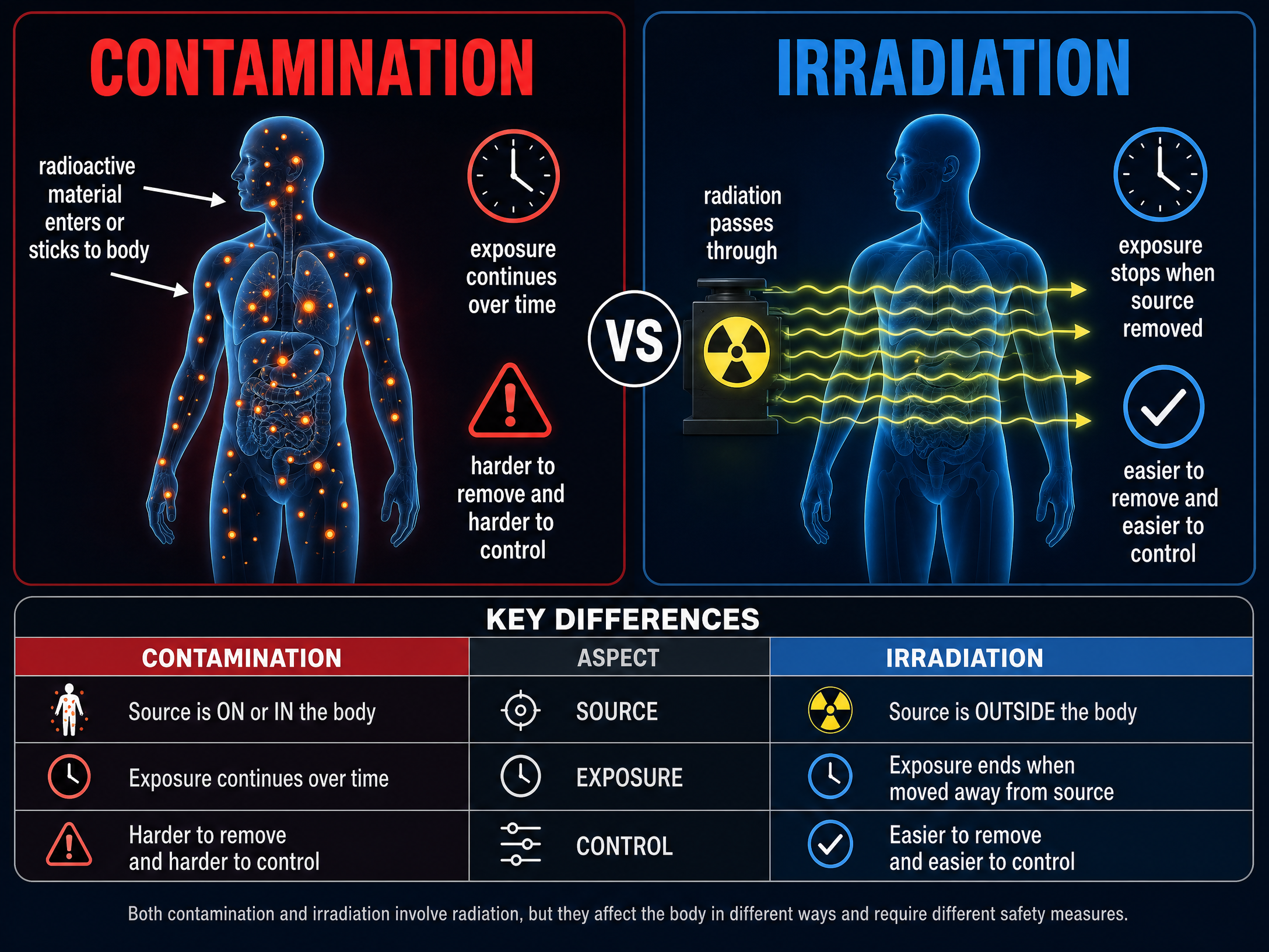
Concept 3: Contamination vs Irradiation

This is one of the most frequently tested distinctions in the specification.
- Irradiation: This is the process of exposing an object to nuclear radiation. The irradiated object does not become radioactive. Exposure stops as soon as the source is removed or shielded. (e.g., getting a medical X-ray).
- Contamination: This is the unwanted presence of materials containing radioactive atoms on other materials. The hazard from contamination is due to the decay of the contaminating atoms. The object remains radioactive as long as the source is on or inside it.
Concept 4: Safety Precautions in Medicine

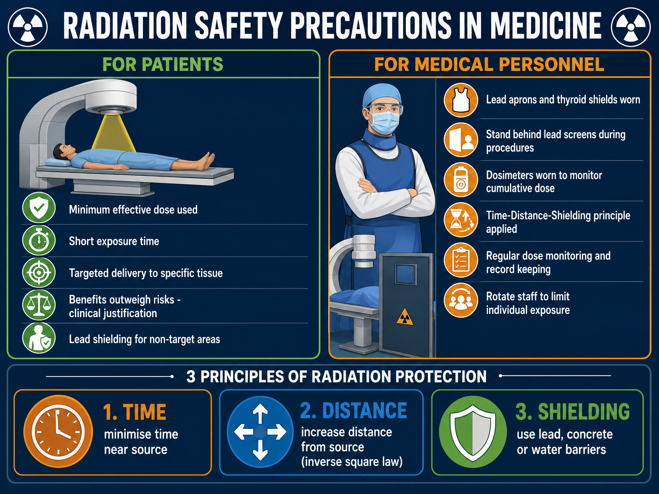
Medical professionals use the principles of Time, Distance, and Shielding to protect themselves and their patients.
- Time: Minimising the time spent near a source reduces the total dose received. Staff may rotate duties to limit individual exposure.
- Distance: Radiation intensity decreases significantly as you move further away (following the inverse square law). Handling sources with long tongs or standing in another room during an X-ray uses distance for protection.
- Shielding: Dense materials absorb radiation. Lead aprons, lead-glass screens, and concrete walls are used to block radiation from reaching healthy tissue.
Mathematical/Scientific Relationships
While this specific sub-topic is largely descriptive, it relies on understanding the properties of the three main types of radiation:
- Alpha (\alpha): Helium nucleus (2 protons, 2 neutrons). Highly ionising, weakly penetrating (stopped by paper/skin).
- Beta (\beta): High-speed electron. Moderately ionising, moderately penetrating (stopped by thin aluminium).
- Gamma (\gamma): Electromagnetic wave. Weakly ionising, highly penetrating (stopped by thick lead/concrete).
Practical Applications
- Radiotherapy: Using high doses of targeted gamma radiation to deliberately cause tissue damage and kill cancer cells.
- Medical Tracers: Injecting or swallowing a gamma-emitting isotope (contamination) so its path through the body can be detected externally. Gamma is used because it can penetrate out of the body and is less ionising than alpha.
- Sterilisation: Medical equipment is irradiated with high-dose gamma rays to kill bacteria. The equipment is irradiated, not contaminated, so it is perfectly safe for surgeons to use.
Visual Resources
3 diagrams and illustrations
Interactive Diagrams
2 interactive diagrams to visualise key concepts
Conceptual Flow Outline
Flowchart showing the decision path for determining radiation hazards based on source location.
Conceptual Flow Outline
The biological pathway from physical ionisation to health outcomes.
Worked Examples
3 worked examples — open one to explore the question and available guidance.
Practice Questions
Test your understanding — click to reveal model answers
State what is meant by the term 'irradiation'. (1 mark)
Hint: Think about whether the object itself becomes radioactive.
A worker in a nuclear power plant accidentally inhales some radioactive dust emitting alpha particles. Explain why this is highly dangerous. (3 marks)
Hint: Think about the properties of alpha radiation and where the source now is.
Compare the hazards of contamination and irradiation. (4 marks)
Hint: Define both, then explain which is generally worse and why.
Explain how ionising radiation can lead to cancer. (3 marks)
Hint: Start at the atomic level and work up to the cellular level.
A hospital uses a gamma-emitting isotope to sterilise plastic syringes. Explain why the syringes do not become radioactive, and why gamma is used instead of alpha. (4 marks)
Hint: Use the terms irradiation and penetration.