OCR · GCSE · Physical Education
Cardiovascular System: Structure, Function, and Response to Exercise
This comprehensive study guide covers the Cardiovascular System (OCR GCSE PE 1.3), focusing on the structure of the heart, the double circulatory system, and the immediate and long-term responses to exercise. Mastering this topic is essential for understanding how the body delivers oxygen to working muscles and adapts to training.
- 8 min read
- 4 worked examples
- 4 practice questions
- 8 key terms
Study Notes
Overview
This guide provides a comprehensive breakdown of the Cardiovascular System, a core component of the OCR GCSE Physical Education specification (1.3). A thorough understanding of the heart's structure, its function, and its immediate and long-term responses to exercise is critical for achieving high marks. Candidates will be expected to apply precise anatomical and physiological knowledge to sporting contexts, analyse data, and evaluate the impact of training on performance.
Key Knowledge & Theory
Core Concepts
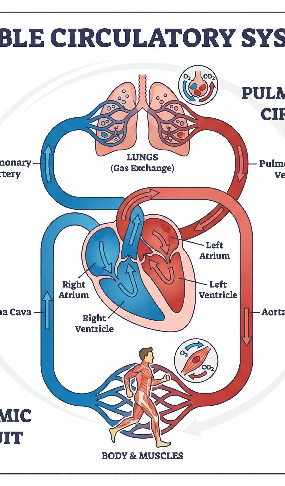
The Double Circulatory System
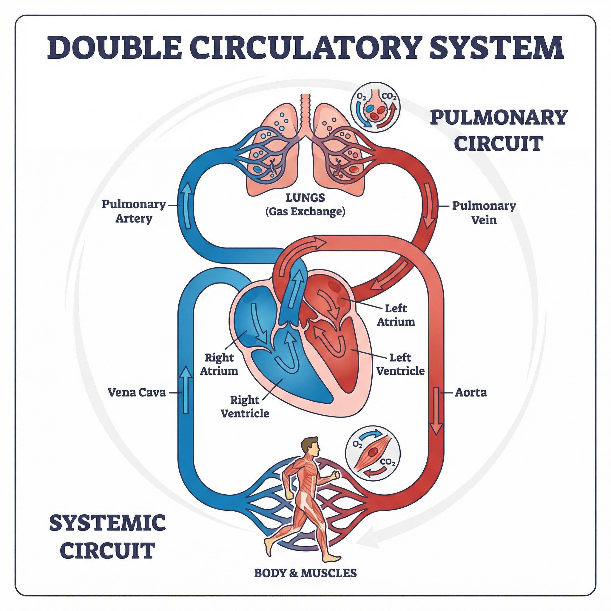
The human cardiovascular system is a double circulatory system, meaning it has two distinct circuits through which blood travels. This is a highly efficient mechanism that ensures oxygenated and deoxygenated blood do not mix, allowing for optimal oxygen delivery to working muscles. Examiners award credit for candidates who can clearly distinguish between these two circuits.
- The Pulmonary Circuit: This circuit is responsible for carrying deoxygenated blood from the right side of the heart to the lungs, and then returning oxygenated blood from the lungs to the left side of the heart. Think Pulmonary = Lungs.
- The Systemic Circuit: This circuit carries oxygenated blood from the left side of the heart to all other parts of the body (muscles, organs) to supply them with the oxygen and nutrients they need for respiration. It then returns the deoxygenated blood back to the right side of the heart. Think Systemic = Body Systems.
The Structure of the Heart
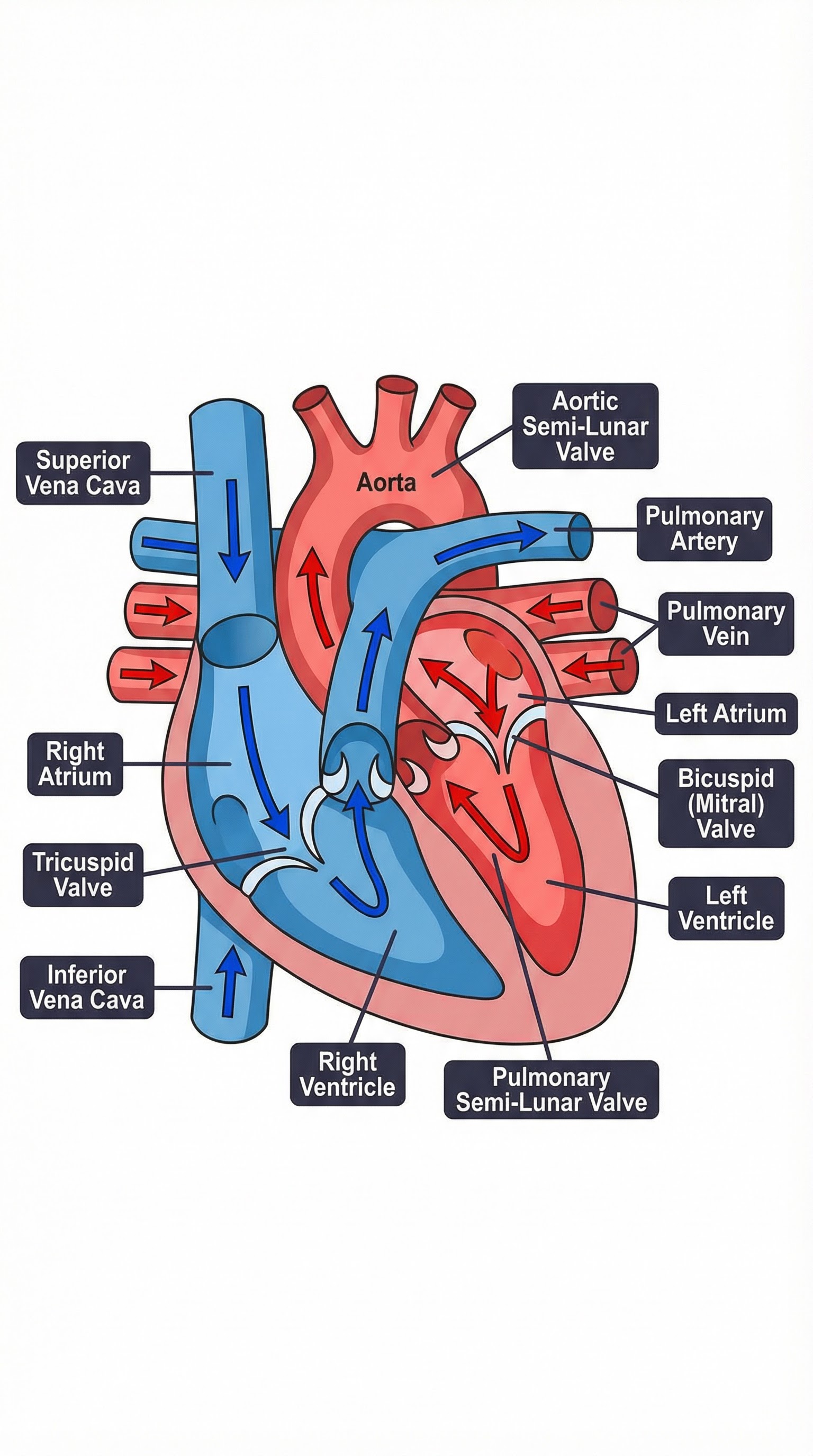
To secure full marks, candidates must demonstrate precise knowledge of the heart's four chambers, the major blood vessels connected to them, and the valves that control blood flow. Using technical terminology is essential.
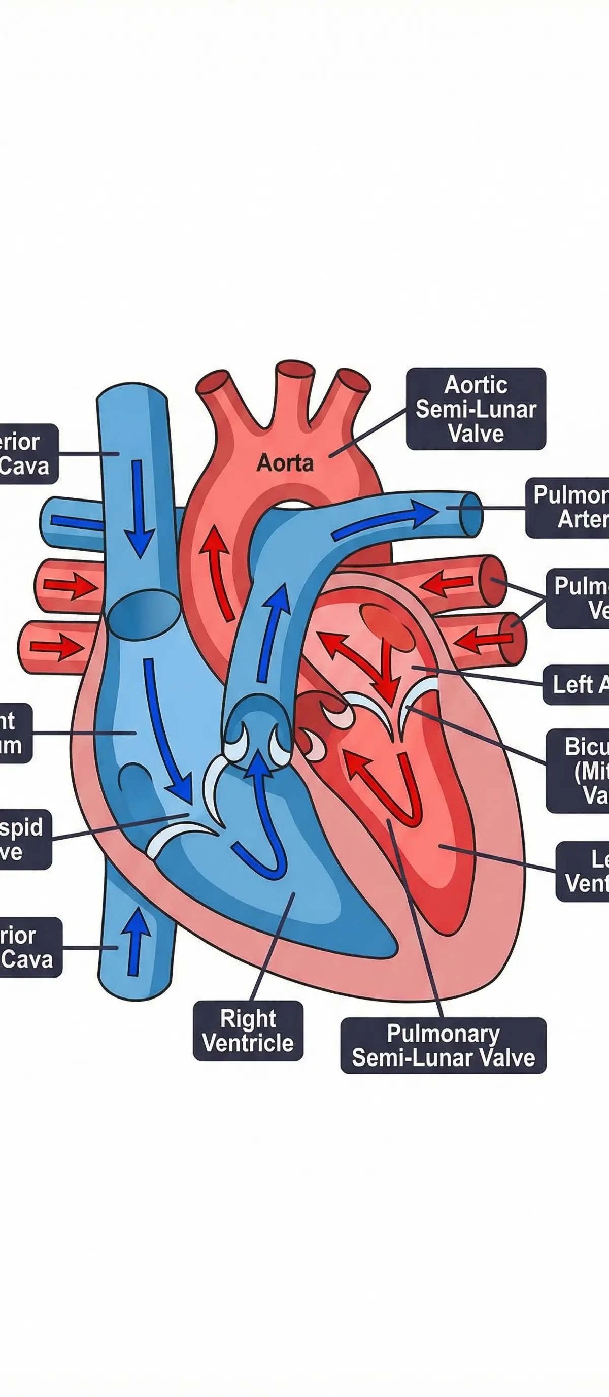
| Chamber/Vessel | Function | Type of Blood |
|---|---|---|
| Vena Cava | The major vein returning deoxygenated blood from the body into the right atrium. | Deoxygenated |
| Right Atrium | Receives deoxygenated blood from the body. | Deoxygenated |
| Tricuspid Valve | Prevents backflow of blood from the right ventricle to the right atrium. | Deoxygenated |
| Right Ventricle | Pumps deoxygenated blood to the lungs via the pulmonary artery. | Deoxygenated |
| Pulmonary Artery | Carries deoxygenated blood from the right ventricle to the lungs. | Deoxygenated |
| Pulmonary Vein | Carries oxygenated blood from the lungs to the left atrium. | Oxygenated |
| Left Atrium | Receives oxygenated blood from the lungs. | Oxygenated |
| Bicuspid (Mitral) Valve | Prevents backflow of blood from the left ventricle to the left atrium. | Oxygenated |
| Left Ventricle | The most powerful chamber; pumps oxygenated blood to the rest of the body via the aorta. Its muscular wall is significantly thicker as a result. | Oxygenated |
| Aorta | The body's largest artery; carries oxygenated blood from the left ventricle to the systemic circuit. | Oxygenated |
| Semi-Lunar Valves | Found in the pulmonary artery and aorta; they prevent blood from flowing back into the ventricles after they have contracted. | Varies |
The Cardiac Cycle: Systole & Diastole
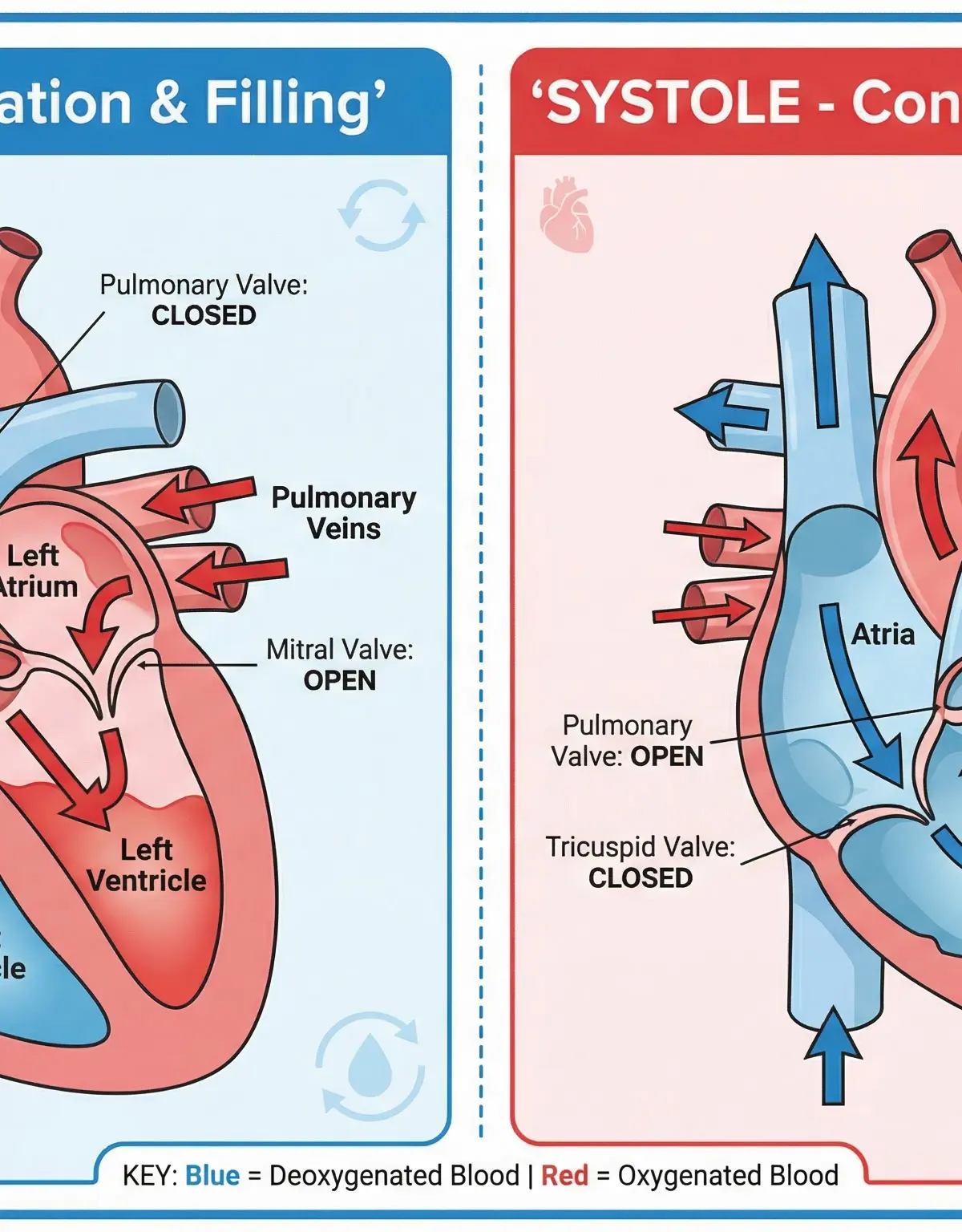
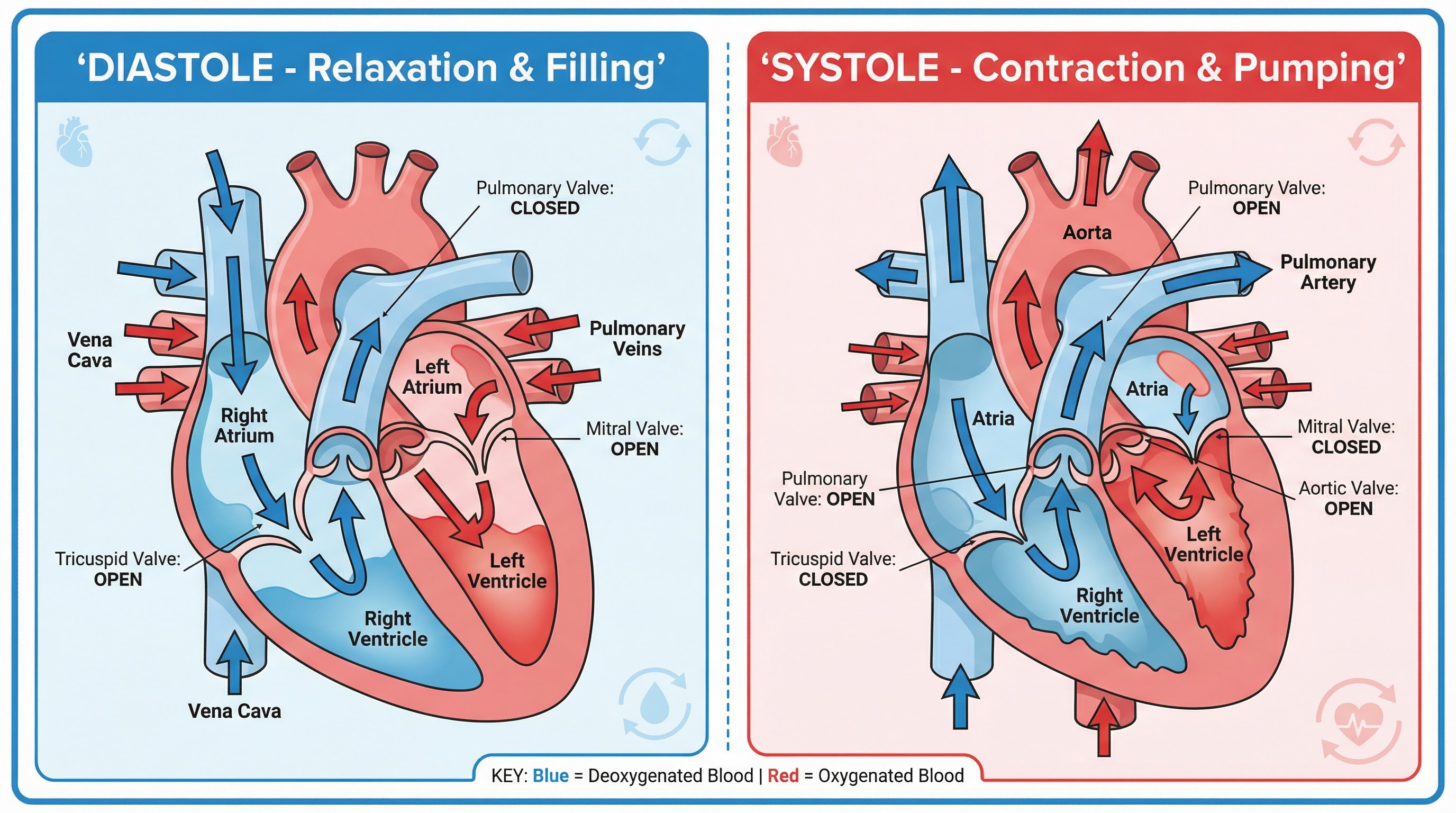
The cardiac cycle describes the sequence of events that occurs during one heartbeat. It consists of two phases: diastole (relaxation) and systole (contraction).
- Diastole: The relaxation phase. The heart chambers fill with blood. The atria contract to push blood into the ventricles, and the tricuspid and bicuspid valves are open.
- Systole: The contraction phase. The ventricles contract forcefully to pump blood out of the heart. The tricuspid and bicuspid valves snap shut (creating the 'lub' sound), and the semi-lunar valves open to allow blood into the aorta and pulmonary artery (the subsequent closing of these valves creates the 'dub' sound).
Technical Vocabulary
Using the correct terminology is a key differentiator between a mid-grade and a top-grade response. You must be fluent in the language of physiology.
- Heart Rate (HR): The number of times the heart beats per minute (bpm).
- Stroke Volume (SV): The volume of blood pumped out of the left ventricle per beat.
- Cardiac Output (Q): The total volume of blood pumped out of the left ventricle per minute. It is the product of heart rate and stroke volume.
- Cardiac Hypertrophy: The thickening and strengthening of the heart muscle (myocardium) as a long-term adaptation to aerobic training.
- Bradycardia: A resting heart rate of below 60 bpm, commonly found in highly trained endurance athletes.
- Vasodilation: The widening of blood vessels to increase blood flow to a specific area (e.g., working muscles during exercise).
- Vasoconstriction: The narrowing of blood vessels to decrease blood flow to a specific area (e.g., non-essential organs during exercise).
Practical Skills
Techniques & Processes: Measuring Cardiovascular Response
In your practical activities, you may be required to measure and analyse the cardiovascular system's response to exercise. A common method is monitoring heart rate before, during, and after exercise.
- Measuring Resting Heart Rate: Before any physical activity, sit in a quiet space for 5 minutes. Place two fingers (index and middle) on your radial artery (at the wrist, in line with the thumb) or carotid artery (in the neck, to the side of the windpipe). Count the number of beats for 30 seconds and multiply by 2 to get your resting HR in bpm.
- Monitoring Exercise HR: During exercise (e.g., on a treadmill or exercise bike), use a heart rate monitor for an accurate reading. Alternatively, stop briefly and take a 15-second pulse count, then multiply by 4. This provides data to show the immediate effects of exercise intensity on the heart.
- Measuring Recovery Rate: After finishing the exercise, record your heart rate every minute for 5-10 minutes. A faster return to resting HR indicates a more efficient cardiovascular system. This is an excellent measure of aerobic fitness.
Exam Component
Written Exam Knowledge
For the written paper, you will be assessed on your ability to recall factual knowledge (AO1), apply it to different scenarios (AO2), and analyse/evaluate the impact on performance (AO3).
The Cardiac Output Equation: Q = SV x HR
This equation is fundamental. You must be able to define each component, state the formula, and use it to calculate missing values from a data set. Marks are awarded for showing your working.
- AO1 (Knowledge): Define Cardiac Output, Stroke Volume, and Heart Rate.
- AO2 (Application): Given that an athlete has a Stroke Volume of 120ml and a Heart Rate of 160bpm during a race, calculate their Cardiac Output in L/min.
- AO3 (Analysis): Explain why an elite marathon runner has a higher cardiac output during exercise compared to an untrained individual.
Short-Term vs. Long-Term Responses
Examiners frequently ask candidates to compare the immediate effects of starting exercise with the long-term adaptations from a sustained training programme.
| Feature | Immediate Response (Starting a 100m sprint) | Long-Term Adaptation (After 6 months of marathon training) |
|---|---|---|
| Heart Rate | Increases dramatically to supply oxygen to muscles. | Resting HR decreases (Bradycardia). Sub-maximal HR is lower. |
| Stroke Volume | Increases as the heart contracts more forcefully. | Resting and exercise SV increases significantly. |
| Cardiac Output | Increases significantly to meet oxygen demand. | Resting Q stays the same, but maximal Q is much higher. |
| Heart Muscle | No immediate change. | Cardiac Hypertrophy occurs; the left ventricular wall becomes thicker and stronger. |
Listen to the Podcast
For an in-depth audio breakdown of these concepts, exam tips, and a quick-fire quiz, listen to our dedicated podcast episode.

Visual Resources
3 diagrams and illustrations
Interactive Diagrams
1 interactive diagram to visualise key concepts
Conceptual Flow Outline
Complete pathway of blood through the double circulatory system
Worked Examples
4 worked examples — open one to explore the question and available guidance.
Practice Questions
Test your understanding — click to reveal model answers
State the equation for Cardiac Output and define each component.
Hint: Think about the three key variables: Q, SV, and HR. What does each letter stand for?
Explain two immediate effects on the cardiovascular system when a sprinter starts a 100m race.
Hint: Think about what happens to heart rate and stroke volume as soon as exercise begins. Why do these changes occur?
A cyclist has a heart rate of 150 bpm and a cardiac output of 24 L/min during a race. Calculate their stroke volume. Show your working.
Hint: Rearrange the equation Q = SV x HR to make SV the subject. Remember to convert units if necessary.
Evaluate the impact of cardiac hypertrophy on the performance of an endurance athlete.
Hint: Think about the chain of adaptations: cardiac hypertrophy → increased stroke volume → bradycardia. How does each of these benefit performance?